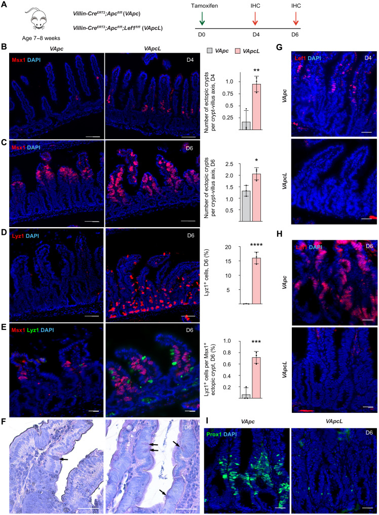
Fig. 6. Lef1 deletion increases the number of ectopic crypts in Apc-mutant adenomas.
(A) Villin-CreERT2;Apcfl/fl (VApc) and Villin-CreERT2;Apcfl/fl;Lef1 fl/fl (VApcL) mice received a single dose of tamoxifen at the age of 7 to 8 weeks, followed by immunohistochemistry analysis of the intestine 4 and 6 days thereafter. (B and C) Msx1 immunostaining and quantification of Msx1+ areas per crypt-villus axis in the intestines (B) 4 days and (C) 6 days after tamoxifen treatment. Scale bars, 50 μm. n = 3 per group, *P < 0.05 and **P < 0.01. (D) Lyz1 immunostaining and quantification of Lyz1+ cells in the intestine on day 6. Scale bars, 50 μm. n = 3 per group, ****P < 0.001. (E) Lyz1 and Msx1 immunostaining and quantification of Lyz1+ cells in Msx1+ ectopic crypt areas on day 6. Scale bars, 50 μm. n = 3 per group, ***P < 0.005. (F) HE (hematoxylin and eosin) images of VApc and VApcL intestines 6 days after tamoxifen. Arrows point to Paneth cells. Scale bars, 100 μm. (G and H) Lef1 immunostaining in the VApc and VApcL intestines (G) 4 days and (H) 6 days after tamoxifen treatment. Scale bars, 50 μm. (I) Prox1 immunostaining in the VApc and VApcL intestines 6 days after tamoxifen treatment. Scale bars, 50 μm. Data are shown as means ± SD. Each dot represents an average value analyzed from individual mouse.

