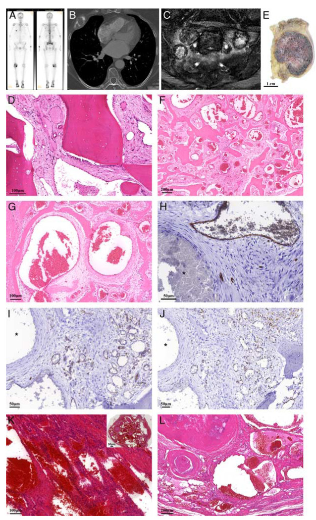
FIGURE 2.
Case 1. A 63-year-old female with 2 benign vascular tumors of bone containing EWSR1-NFATC2 fusion. A, Bone scintigraphy with increased uptake of the tracer in the lesion in the rib, but also, focal abnormalities in the skull, spine and sacrum. B, CT image of the chest with an expansile osteolytic lesion in the fifth rib on the right side. C, Coronal T2-weighted MR image with fat suppression. Multiple lesions in the sacrum with variable signal intensities from predominantly low, as the one in the center, to mixed low and higher and high. Surrounding perilesional edema. D, Hematoxylin and eosin staining of biopsy of the sacral mass shows a tumor mainly composed of thin-walled vessels with no atypia of the lining cells. E, Gross specimen of the rib lesion showing a well-demarcated red tumor in the medullary cavity of the ventral part of the rib with cortical expansion. Low (F) and higher (G) power view of hematoxylin and eosin staining showing that the rib tumor is mainly composed of large dilated blood vessels of variable caliber, some of which are thin walled, while others have a thicker wall, lying within preexisting trabecular bone. Vascular markers CD34 (H), CD31 (I), and ERG (J) confirmed the endothelial lining in only a subset of the spaces, while some of the cysts containing a thicker wall are negative (asterix). Note the background of loose myxoid to more fibrous stroma with bland stromal cells. K and L, Hematoxylin and eosin staining of the vascular lesion in the soft tissue of her foot 22 years previously demonstrating a sharply defined lesion (inset) with different morphology containing mainly numerous large dilated, thin-walled vessels containing prominent phleboliths, admixed with focal spindle cell areas.

