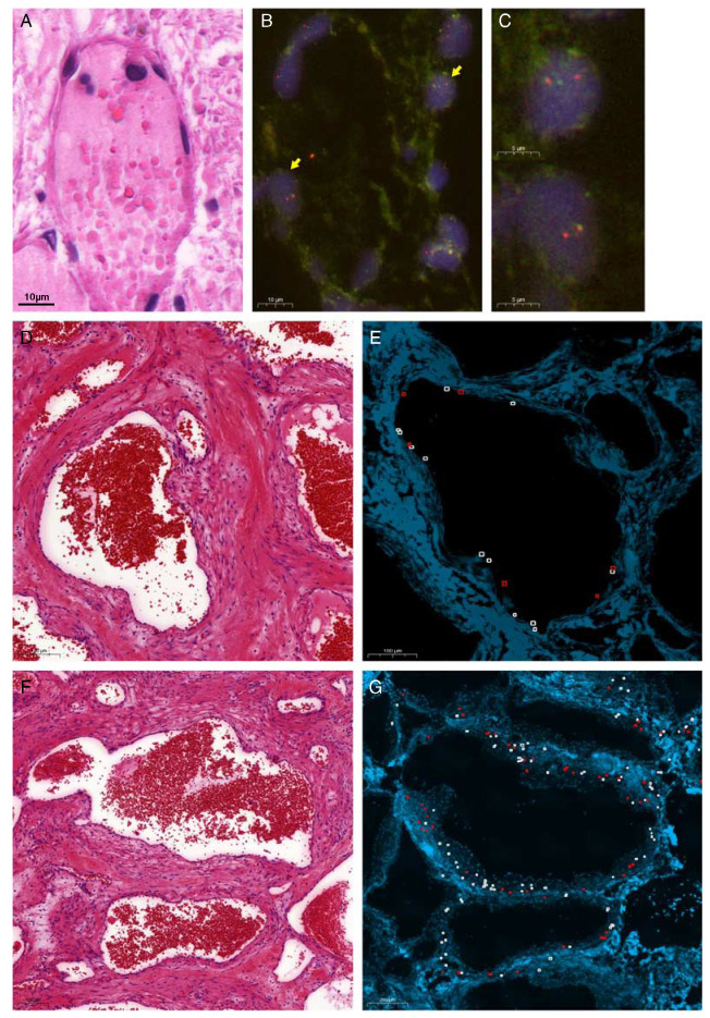
FIGURE 5.
EWSR1-NFATC2 translocation identified in the endothelial lining as well as the surrounding spindle cells in vascular malformation/hemangioma. EWSR1-NFATC2 colocalization FISH (B, C) and corresponding area in hematoxylin and eosin section (A) in L6952 (case 1) highlights the presence of the fusion in the endothelial lining. Yellow arrows marking fusion cells. Scoring of EWSR1-NFATC2 FISH in case 2 for the lining cells (D, E) and the surrounding spindle cells (F, G). Red boxes represent cells with colocalization of the EWSR1 and NFATC2 probe, while white boxes represent normal cells.

