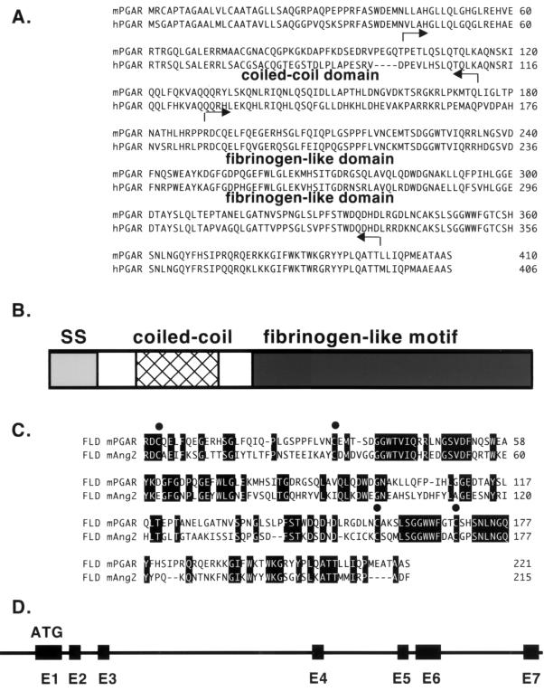
FIG. 1.
Sequence analysis of PGAR. (A) Deduced amino acid sequences of mouse PGAR (mPGAR) and human PGAR (hPGAR). The arrows indicate the limits of the coiled-coil and fibrinogen-like domains. (B) Schematic diagram of the predicted PGAR protein structure. (C) Alignment of the fibrinogen-like domains (FLD) of PGAR and angiopoietin-2 (Ang2). Conserved cysteines are indicated by solid circles. SS, signal sequence. (D) Genomic structure of PGAR. Exons are denoted by black boxes, and introns are denoted by a solid line.

