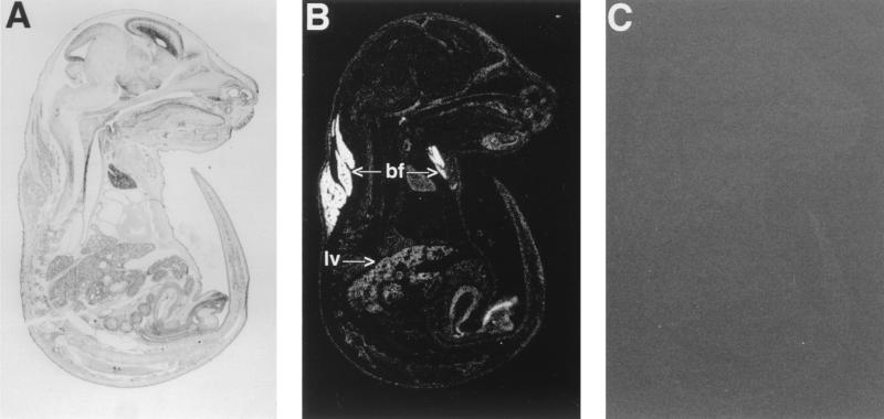
FIG. 4.
In situ analysis of PGAR in mouse. Shown are parasagittal sections (8 μm thick) of E18.5 mouse embryo hybridized to antisense (B) or sense (C) PGAR RNA probe. Arrows point to subscapular brown fat (bf) and liver (lv). Also shown is a bright-field image of a hematoxylin-and-eosin-stained section (A).

