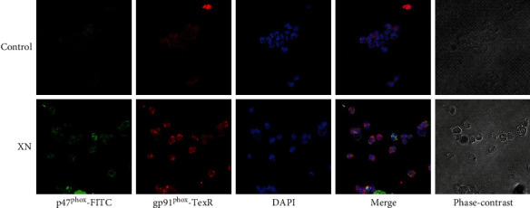
Figure 3.

Representative images showing XN-induced colocalization between NOX subunits gp91phox and p47phox. After treated with 10 μM XN for 1 h, cells were collected, washed, fixed, and incubated with gp91phox and p47phox primary antibodies overnight at 4°C and then probed with TR and FITC conjugated secondary antibodies, respectively. The fluorescent images were obtained with a confocal microscope (200x).
