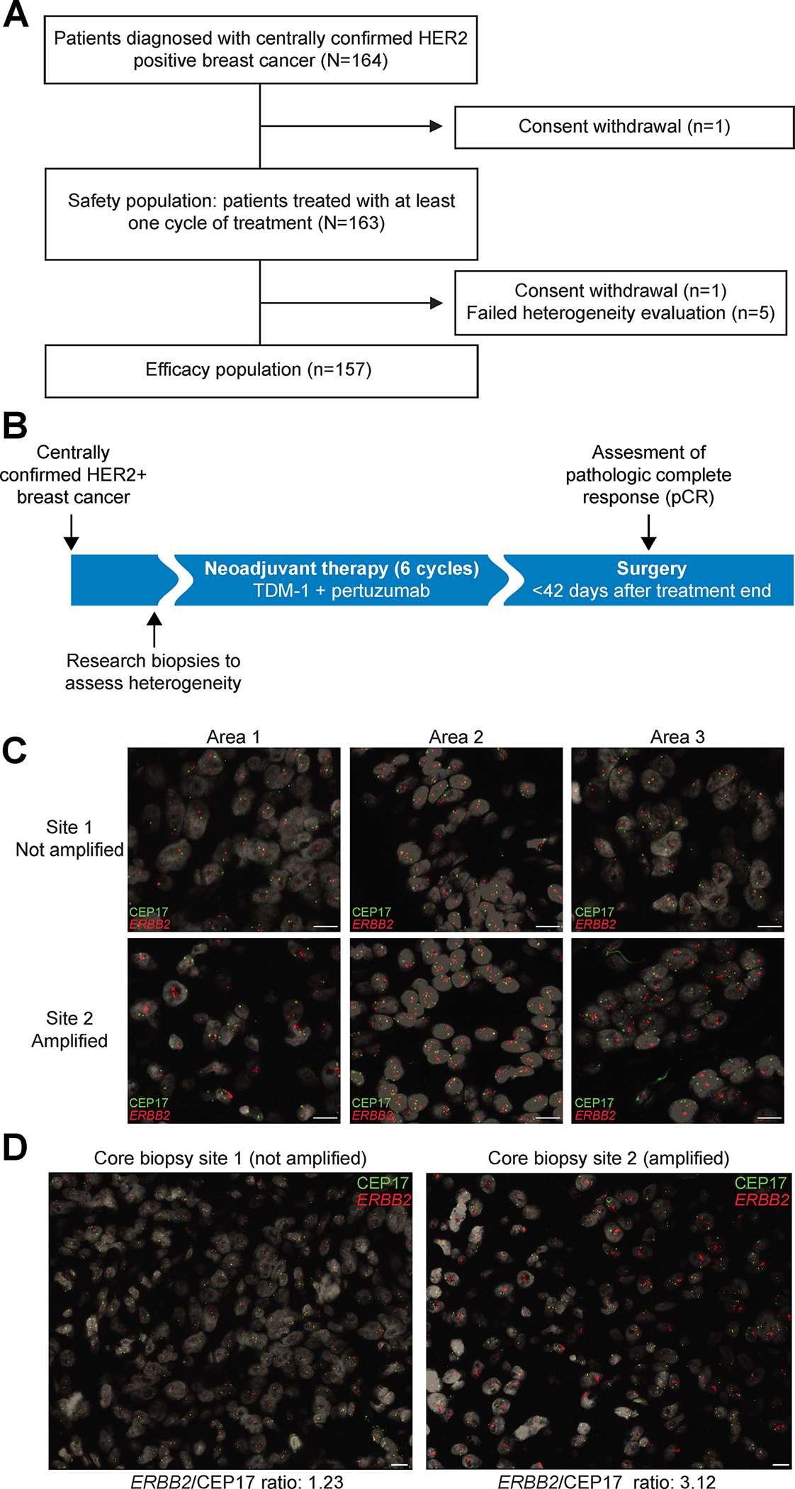
Figure 1. Study Design.

A, CONSORT diagram. B, Study design. Centrally confirmed HER2-positive breast cancer patients were treated in a single arm study. Treatment consisted of six infusions of T-DM1 given in combination with pertuzumab. C, Example of central pathology evaluation of HER2 heterogeneity assessed by FISH, with CEP17 probe in green and ERBB2 in red. ERBB2 copy number counting was performed in three different areas per core biopsy site counting approximately 50 cells in each area. Scale bar corresponds to 10 μm. D, A representative case of HER2 heterogeneity with core biopsy site 1 showing HER2-negative cancer and core biopsy site 2 HER2-amplified cancer. Scale bar corresponds to 10 μm.
