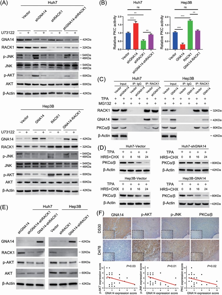
Figure 6.
GNA14 inhibits the activity of AKT and PKC induced by RACK1. (A) The protein level of GNA14, p-JNK and p-AKT were detected by western blot using indicated antibodies in GNA14- and/or RACK1-intervened Huh7 and Hep3B cell lines after treating with or without U73122 (10 μM, 30 min). (B) PKC activity of GNA14- and/or RACK1-intervened Huh7 and Hep3B cell lines were measured by using a non-radioactive protein kinase C assay kit. Relative PKC activity in Huh7-Vector and Hep3B-Vector were taken as a control of 100%. Data shown are from three experiments. (C) Huh7, Hep3B and their lentivirus-infected cells were treated with TPA (200 nM) and MG132 (20 μM) for 30 min. Treated cells were collected, lysed, and used for RACK1 immunoprecipitation, followed by western blot analysis using the indicated antibodies. (D) Huh7, Hep3B and their lentivirus-infected cells were treated with TPA (200 nM, 30 min) and cycloheximide (10 μg/ml) at different time points as indicated followed by western blot analysis using the indicated antibodies. (E) The protein level of GNA14, RACK1 and p-AKT were detected by western blot using indicated antibodies in GNA14- and/or RACK1-intervened Huh7 and Hep3B cell lines. (F) Representative IHC images of GNA14, p-AKT, p-JNK and PKCα/β protein level in HCC serial slices. magnification: 100×, inset magnification: 400×. The correlation analysis between GNA14 and p-AKT, GNA14 and p-JNK, GNA14 and PKCα/β were shown in the diagram below.

