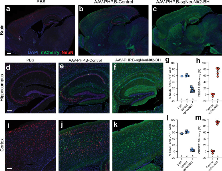
Fig. 5. Immunohistochemical analysis confirms disruption of NeuN in the brain.
a–c General NeuN signal reduction is detected in the hippocampus and the cortex. d–f AAV-PHP.B-sgNeuN#2-BH led to a decrease in NeuN in the CA1 (Cornu Ammonis), CA2 and CA3 regions of the hippocampus. g, h Quantification analysis showing robust CRISPR knockdown efficiency in hippocampal neurons. i–k Sharp reduction of NeuN expression was detected in the superficial and deep regions of the cortex. l, m AAV-PHP.B-sgNeuN#2-BH achieves a high CRISPR efficiency in the cortex. Brain tissues were collected and fixed for double-immuno-staining with anti-NeuN (red) and anti-mCherry (green) antibodies. Data represent means ± SD of n samples noted above the x-axis. Scale bars: (a–c) = 500 μm, (d–f) = 200 μm, (i–k) = 100 μm. (color figure online).

