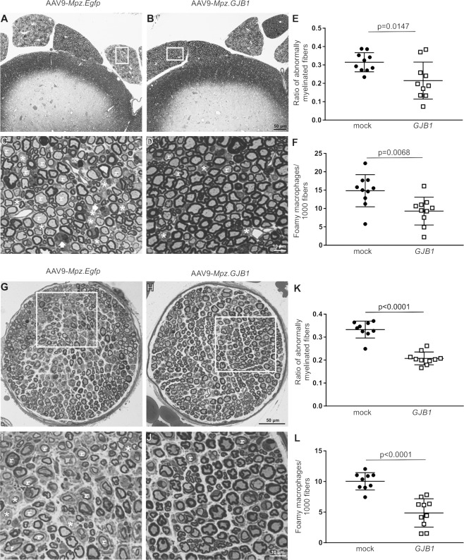
Fig. 5. Morphological analysis of anterior lumbar roots and femoral motor nerves of Gjb1-null mice following post-onset intrathecal delivery of the AAV9-Mpz.GJB1 vector.
These are representative images of semithin sections of anterior lumbar spinal roots attached to the spinal cord at low (A) and (B) and higher (C) and (D) magnification, with morphometric analysis results (E) and (F), as well as of semithin sections of femoral motor nerves at low (G) and (H) and higher (I) and (J) magnification with related morphometric analysis results (K) and (L), from mock and full (GJB1) vector treated mice as indicated, at 10 months of age (4 months after treatment). AAV9-Mpz.GJB1 injected mice show improved myelination compared with mock-treated littermates with fewer demyelinated (*) and remyelinated (r) fibers in both tissues as well as fewer foamy macrophages (arrows in C). Quantification of the ratios of abnormally myelinated fibers in multiple roots and femoral nerves (n = 10 mice per group) confirms significant improvement in the numbers of abnormally myelinated fibers (E) and (K) as well as significant reduction in the numbers of foamy macrophages (F, L) in the treated compared with mock treated littermates (see also Tables S4 and S6). Scale bars: (A, B), (G, H) 50 μm, (C, D), (I, J) 10 μm.

