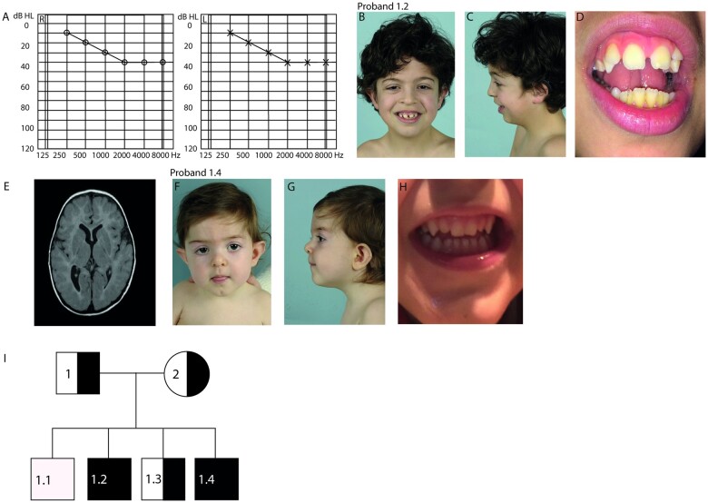
Figure 1.
NAA80 individuals present with craniofacial dysmorphisms, abnormal brain MRI and high-frequency hearing loss. (A) Pure Tone Audiometry showing intensity measured in decibels (dB) represented on the x-axis and the frequency measured in Hertz (Hz) on the y-axis of the left ear (L) and right ear (R) of proband 1.2 showing high frequency hearing loss in the 2000–8000 kHz region. (B) Front view of proband 1.2 at the age of 7 years showing small upper lip, hypertelorism, abnormally shaped ears, ptosis and epicanthus fold. (C) Lateral view of proband 1.2 showing retrognathia. (D) Brain MRI image of proband 1.2 showing asymmetrical posterior horns. (E) Close-up of proband 1.2 showing diastema and peg-shaped lateral incisors. (F) Front view of proband 1.4 at the age of 1 year showing small upper lip, long philtrum, bulbous tip of the nose, ptosis, epicanthus fold, ocular hypertelorism, abnormally shaped ears. (G) Lateral view of proband 1.4. (H) Close-up of proband 1.4 showing peg-shaped lateral incisors. (I) Family pedigree of the NAA80 family. Black squares indicate affected male [homozygous for NAA80 c.389T>C, p.(Leu130Pro) variant]. Semi-black squares indicate that the individual is a carrier of a heterozygous NAA80 variant.

