
Fig. 3.
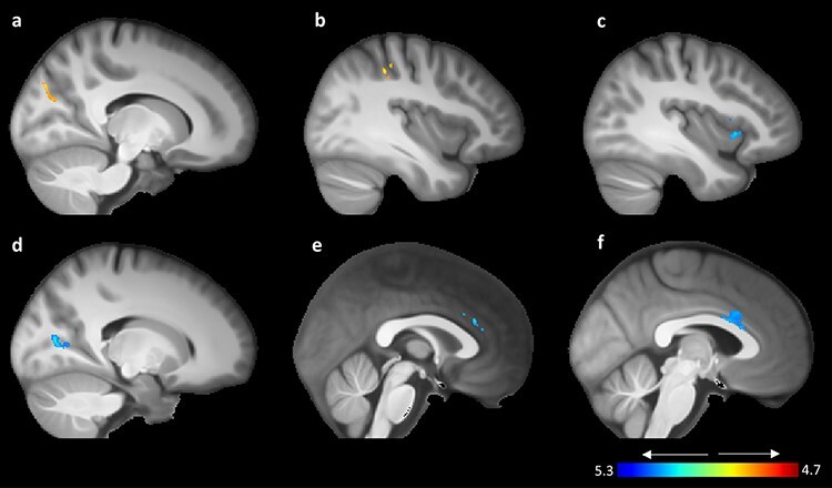
VBM GMV findings for positive (red–yellow color bar) and negative (blue–light blue color bar). All findings are overlaid on a normalized averaged skull-extracted brain template created from the participant cohort and presented from a sagittal view. (a) Right medial visual association area, (b) right intraparietal sulcus, (c) right insula, (d) right primary visual cortex, (e and f) dACC.