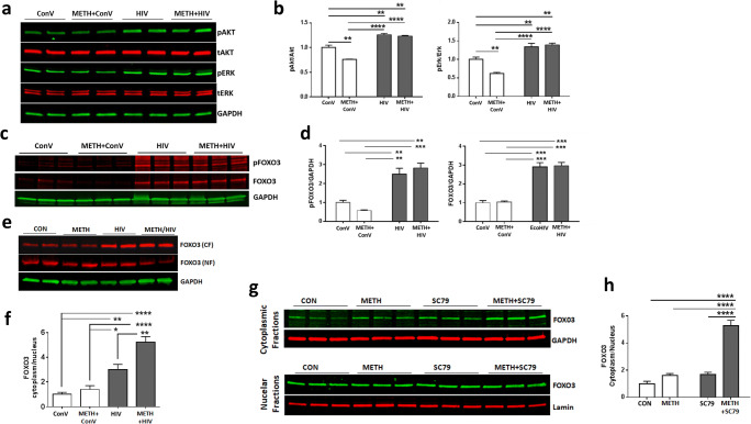
Fig. 6.
METH exposure and HIV infection enhance the sequestration of FOXO3 in the cytoplasm. a METH exposure and/or HIV infection-induced activation of Akt and Erk. ReNcells were exposed to 100 μM METH for 24 h, followed by infection with HIV (60 ng/ml of p24) for 48 h. Cell lysates were separated on SDS-PAGE to evaluate the expression of phosphorylated and total Akt (pAkt and tAkt, respectively) as well as phosphorylated and total Erk (pErk and tErk, respectively). Representative images are presented. GAPDH was used as a loading control. b Quantitative results from (a). Two-way ANOVA, N=6 per group. **, p<0.01, and ****, p<0.001. c METH exposure and/or HIV infection-induced protein levels of phosphorylated FOXO3 (pFOXO3) and total FOXO3. Representative immunoblots. d Quantitative results from c. Two-way ANOVA, N=3 per group. **, p<0.01, and ***, p<0.001. e Representative images of FOXO3 in the cytoplasmic (CF) and nuclear (NF) fractions. FOXO3 protein levels were normalized to the GAPDH levels. f The ratio of FOXO3 in the cytoplasmic to nuclear fractions from (e). N=6 per group. *, p<0.05, **, p<0.01, and ****, p<0.001. g Effect of Akt activator SC79 on FOXO3 subcellular localization. ReNcells were exposed to 100 μM of METH for 24 h and twice treated with 5 μM SC79 in a 24 h interval. Cells were harvested 24 h after the second treatment with SC79 and separated into the cytoplasmic and nuclear fractions. GAPDH and Lamin A were used as loading controls for the cytoplasmic and nuclear fractions, respectively. h The ratio of FOXO3 in the cytoplasmic to nuclear fractions from (g). N=6 per group. ****, p<0.0001

