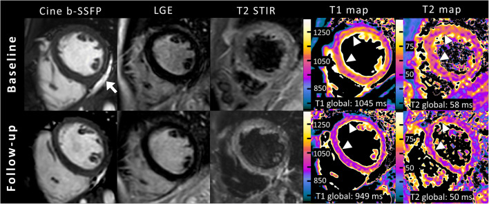
Fig. 5.
A clinical example of cardiac magnetic resonance imaging (MRI) in short-axis view in a 16-year-old boy. Cine images (balanced steady state free precession [b-SSFP]) show normal left ventricular ejection fraction (LVEF; 58%, no segmental hypokinesia) and pericardial effusion basal inferior (arrow). No focal or diffuse enhancement was identified on late gadolinium enhancement (LGE). No focal myocardial edema was visible on fat-suppressed (T2-weighted short TI inversion recovery [T2 STIR]) images. Mapping parameters displayed high global myocardial native T1 and T2 relaxation times at baseline cardiac MRI and normalization at follow-up (arrowheads show the most affected segments). The diagnosis of acute diffuse myocarditis in this patient was only possible using quantitative parameters according to 2018 Lake Louise criteria and would have been missed by the original Lake Louise criteria

