Fig. 4.
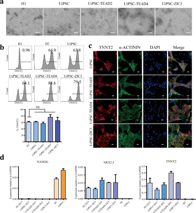
Induce UiPSCs-TEAD2, UiPSCs-TEAD4, and UiPSCs-ZIC3 differentiate to myocardial cells. a The cell morphology of induced myocardial cells from H1, UiPSCs, UiPSCs-TEAD2, UiPSCs-TEAD4 and UiPSCs-ZIC3 (bar: 200 μm). b FACS assay to detect the proportion of TNNT2+ cells after 15 days myocardial cells differentiation. c Immunofluorescence experiment to detect the expression of myocardial markers TNNT2 and α-actinin in differentiated myocardial cells. The nuclei were stained with DAPI (bar: 20 μm). d qPCR was used to detect the expression of pluripotency markers (NANOG) and myocardial markers (NKX2.5 and TNNT2) in differentiated cells and pluripotent cells. Data represented as mean ± SEM from three independent assays. *P < 0.05, **P < 0.01, ***P < 0.001, unpaired two tailed student-t-test

