Fig. 6.
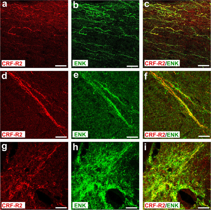
Double immunofluorescence staining of CRF-R2 (a, d, g) and ENK (b, e, h) in the rat L4-L5 spinal cord. (a, b, c) Parasagittal sections of L4-L5 spinal cord of the rat show a network of CRF-R2-immunoreactive fibers overlapping with ENK and extending through the superficial laminae of the dorsal horn of the lumber spinal cord. Some fibers express only ENK. (d–i) Double immunofluorescence staining of coronal sections of spinal cord of the rat showing that CRF-R2-immunoreactive fibers overlap with ENK. Some fibers express ENK only. Bar = 20 μm

