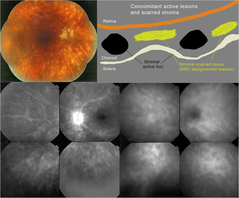
Fig. 19.
HLA-A-29 BRC. The typical fundal cream coloured rice shaped lesions are shown on the fundus picture (top left). The marked retinal vasculitis is shown on FA in the left bottom quartet. The choroidal foci are shown on ICGA in the right bottom quartet. The cartoon (inspired by Joan Mirò) (top right) explains that the cream-coloured lesions (yellow) do not correspond to the ICGA HDDs and do not appear on ICGA. HDDs correspond to the black dots on the cartoon

