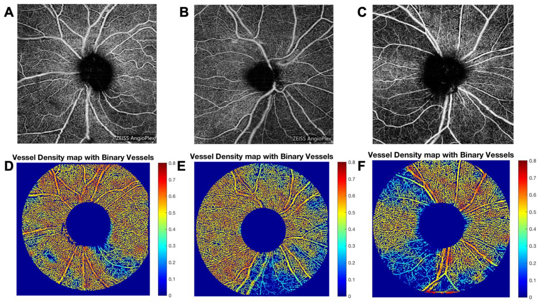
Figure 1: 6×6-mm OCTA en face images centered on the ONH of (A) mild, (B) moderate, and (C) severe POAG. Corresponding vessel density map with binary vessels of (D) mild, (E) moderate, and (F) severe POAG.

Areas of higher vessel density are in warmer colors. Abbreviations: OCTA=optical coherence tomography angiography; ONH=optic nerve head; POAG=primary open angle glaucoma.
