
Figure 2.
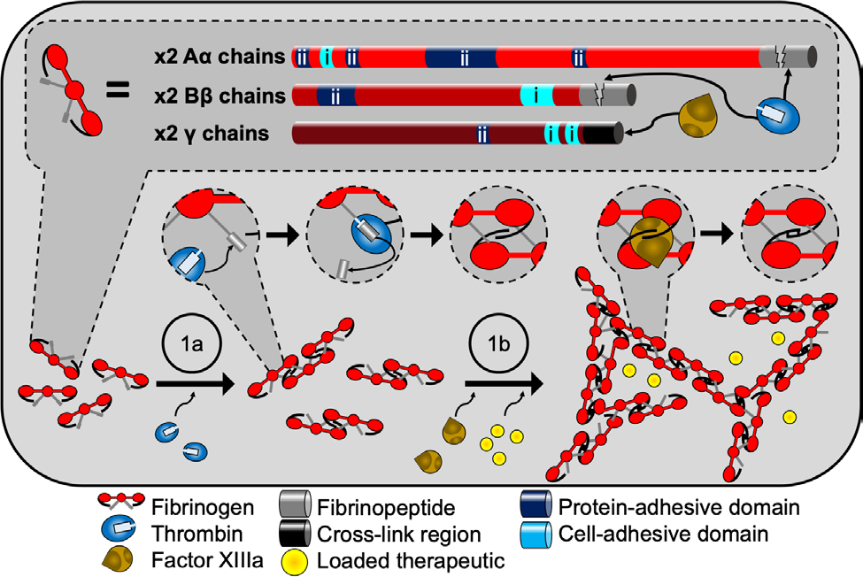
Fibrin hydrogels provide multiple bioactive interfaces. Fibrinogen is composed of two sets of three peptide chains called Aα Bβ, and γ (top). On each chain exists various binding sites for both cells (i) and several ECM proteins (ii). Fibrin hydrogel formation (bottom) is most often carried out in a batch solution containing fibrinogen, calcium, and the enzymes thrombin and factor XIIIa. First (1a), thrombin cleaves off the fibrinopeptides on chains Aα and Bβ which initiates polymerization and fibril formation via intermolecular association between the cleaved site of one molecule and a binding site from another. Next (1b), factor XIIIa covalently links the fibrinogen units together via overlapping γ chains. If therapeutics are used, they are included in the batch mixture before gelation.