
Figure 2. Co-crystal structure of rearranged during transfection (RET)- tyrosine kinase inhibitor (TKI) complex.
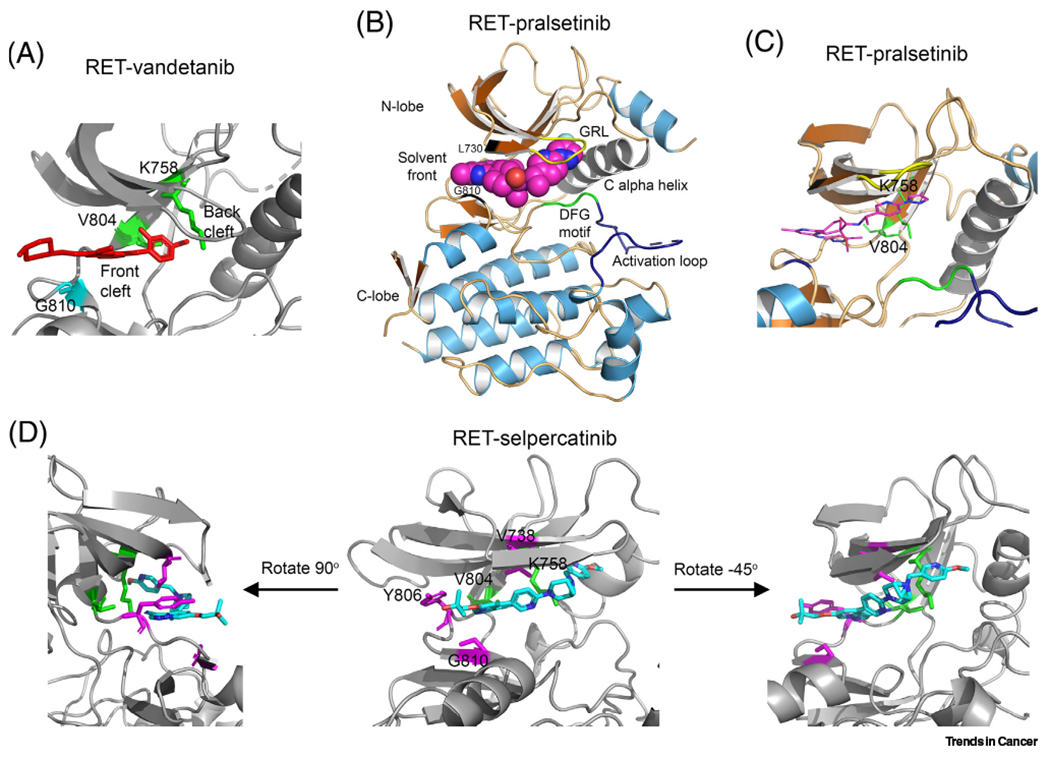
(A) RET-vandetanib complex [Protein Data Bank (PDB): 2IVU] [82]. Red, vandetanib; green, V804 gatekeeper residue and K758 gatewall residue; cyans, G810 solvent front residue. (B,C) Co-crystal structure of RET–pralsetinib complex (PDB: 7JU5) [83]. (D) Co-crystal structure of RET-selpercatinib complex (PDB: 7JU6) [83]. Gatekeeper V804 and gatewall K758 are in green. Magentas denotes residues where selpercatinib-resistant mutations have been identified. Abbreviation: GRL, Gly-rich loop.