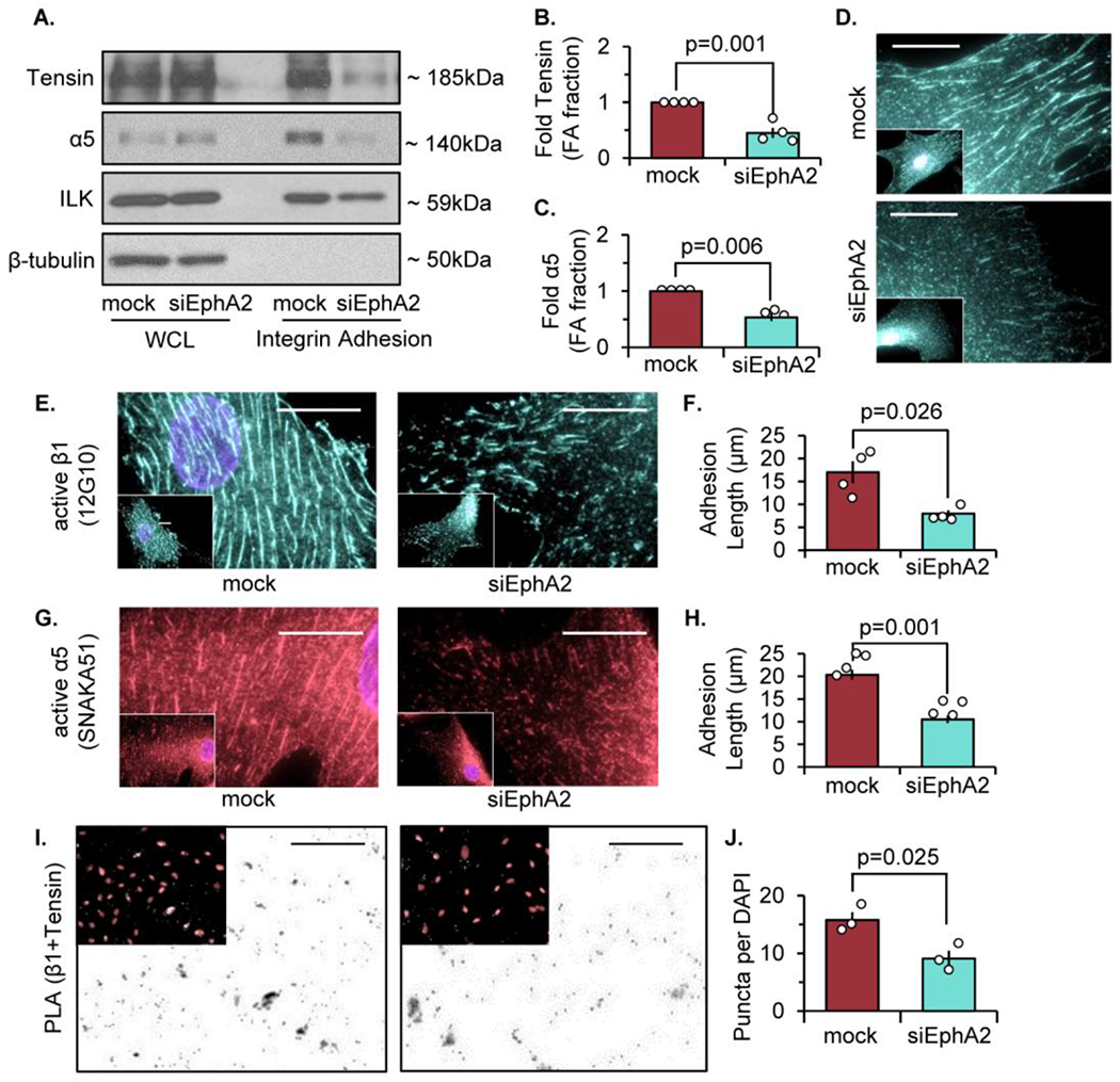
Figure 1: EphA2 depletion reduces tensin localization and fibrillar adhesion length.

Human vascular smooth muscle cells (VSMCs) were transfected with either mock or siRNA targeted against EphA2 for 24 hours. A-C) Cells were plated onto Matrigel-coated glass slides overnight in 1% serum, and integrin adhesion isolation was performed. Protein expression was measured by immunoblot and normalized to integrin-linked kinase (ILK). β-tubulin from the whole cell lysate (WCL) fraction was shown for integrin adhesion isolation purity. B,C) Tensin and α5 integrin from the integrin adhesion fraction were quantified. D) Cells were stained for tensin. E,F) Cells were stained for active β1 integrin (12G10) and adhesion length was quantified in microns. G,H) Cells were stained for active α5 integrin (SNAKA51) and adhesion length was quantified in microns. (I) Proximity ligation assay (PLA) was performed for β1 and tensin interactions, and counterstained with DAPI (pink). Scale bar = 25μm. J) PLA puncta were quantified per high powered field and normalized to number of DAPI per high powered field. Scale bar = 25um. n=3-4. Data are expressed as mean ±SEM. Statistical comparisons were made using Student’s T-test (B,F,H,J,L). A p-value less than 0.05 is considered significant.
