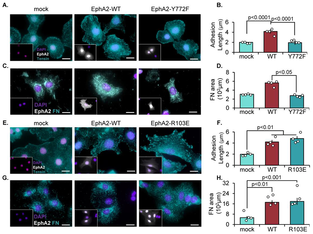
Figure 3: EphA2-dependent fibrillar adhesion elongation requires Y772 phosphorylation but not EphA2-ligand interactions.

EphA2 KO mouse aortic VSMCs were transfected with either mock, EphA2-WT, (A-D) EphA2-Y772F, or (E-H) EphA2-R103E constructs for 24 hours, then plated onto Matrigel-coated coverslips overnight in 1% serum. Cells were stained for EphA2 (white), and EphA2-positive cells were quantified. A-B,E-F) Cells were stained fortensin (teal) and adhesion length was measured in microns. C-D,G-H) Cells were stained for fibronectin (teal) and quantified as fibronectin-positive area in microns. Scale bar = 25μm. n=4-5. Data are expressed as mean ±SEM. Statistical comparisons were made using One-way ANOVA with Bonferroni post-test. A p-value less than 0.05 is considered significant.
