
Figure 6: EphA2 signals through RhoA to promote fibrillar adhesion elongation and fibronectin deposition.
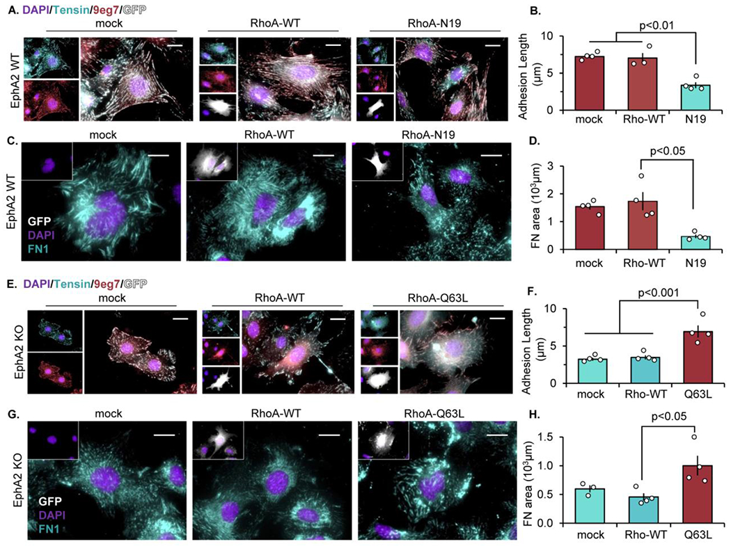
A-D) EphA2 WT mouse VSMCs were transfected with either mock, RhoA-WT, or dominant-negative RhoA (RhoA-N19) for 24 hours. (E-H) EphA2 KO mouse VSMCs were transfected with mock, RhoA-WT, or constitutively active RhoA (RhoA-Q63L) for 24 hours. Cells were plated onto Matrigel overnight post-transfection, and GFP-positive cells (white) were quantified. A/B,E/F) Cells were stained for tensin (teal), active β1 integrin (9eg7, pink), and DAPI (purple) and fibrillar adhesion length was measured in microns. C/D,G/H) Cells were stained for fibronectin (teal) and DAPI (purple) and fibronectin area was measured in microns. Scale bar = 25μm. n=4. Data are expressed as mean ±SEM. Statistical comparisons were made using One-way ANOVA with Bonferroni post-test A p-value less than 0.05 is considered significant.