
Figure 7: Deletion of EphA2 enhances p190Rho-GAP phosphorylation to inhibit fibronectin fibrillogenesis.
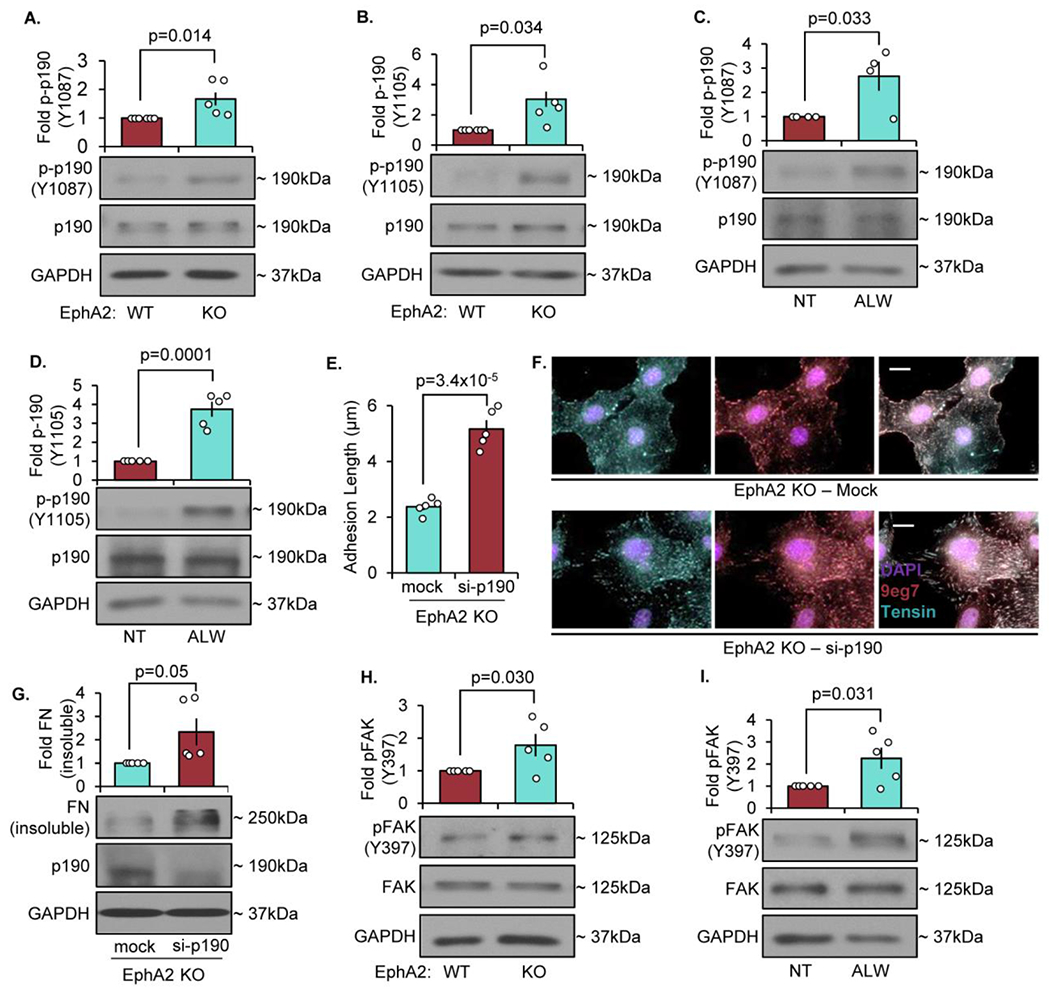
A/B) EphA2 WT or EphA2 KO mouse VSMCs were plated onto Matrigel overnight in 1% serum, and phospho-p190RhoGAP was quantified by Western blot and normalized to total p190RhoGAP. C/D) EphA2 WT mouse VSMCs were plated onto Matrigel overnight in 1% serum, then treated with ALW-II-41-27 (0.5μM) for 30 minutes. Phospho-p190RhoGAP was quantified by Western blot and normalized to total p190RhoGAP. E-G) EphA2 KO mouse VSMCs were transfected with either mock or siRNA against p190Rho-GAP for 24 hours, then plated onto Matrigel overnight. G,H) Cells were stained for tensin (teal), active β1 integrin (9eg7, pink), and DAPI (purple), and fibrillar adhesion length was measured in microns. Scale bar = 25μm. n=4-5. G) Cells were analyzed for fibronectin deposition with deoxycholate extraction. Deoxycholate-insoluble (deposited) fibronectin was normalized to deoxycholate-soluble GAPDH. H/I) EphA2 WT or EphA2 KO mouse VSMCs were plated onto Matrigel overnight in 1% serum, and phospho-FAK Y397 was quantified by Western blot and normalized to total FAK. Data are expressed as mean ±SEM. Statistical comparisons were made using Student’s T-test. A p-value less than 0.05 is considered significant.