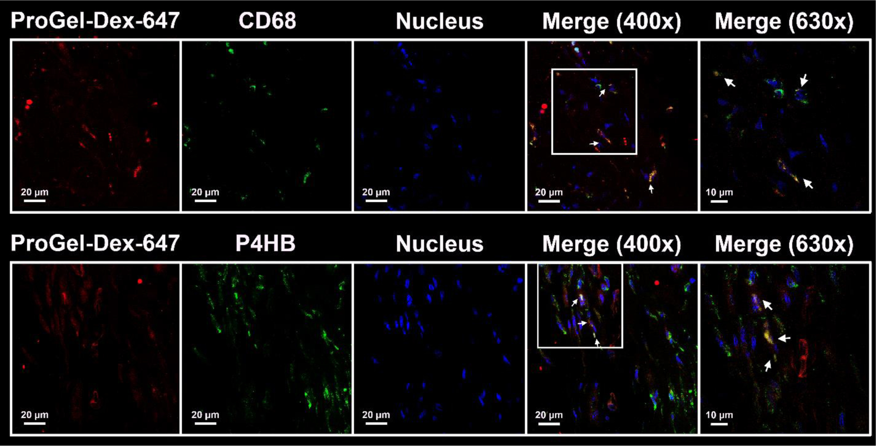
Figure 5.

Representative confocal images of anti-CD68 and anti-P4HB antibody stained sections of decalcified MAA rat knee joints. Each panel is composed of five subpanels: Anti-CD68 or anti-P4HB signal (green), Alexa 647-labeled ProGel-Dex (red), DAPI signal (blue), and two merged images at 400× and 630× magnification. The colocalization of the red and green colors confirmed the internalization of the Alexa 647-labeled ProGel-Dex by P4HB-positive cells (fibroblasts) and CD68-positive cells (monocytes/macrophages) of the synovial lining. Arrows point to the colocalization of cell markers and Alexa 647-labeled ProGel-Dex.
