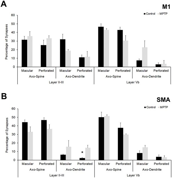
Figure 5 .

Quantitative analysis of PSDs (perforated vs. macular) of synapses formed by vGluT2-immunopositive synapses in superficial and deep layers in control and parkinsonian animals. (A) Percentage of axo-dendritic and axo-spine synapses with macular or perforated PSDs in cortical layers of M1. (B) Analysis of macular and perforated PSDs in cortical layers of SMA. Except for perforated axo-dendritic synapses in layer II–III of SMA (B), more frequently encountered in MPTP-treated monkeys than in controls (Student’s t-test, *P = 0.007), there are not statistically significant differences in the percentage of macular versus perforated PSDs between control and parkinsonian monkeys in both M1 (A) and SMA (B).
