Figure 1.
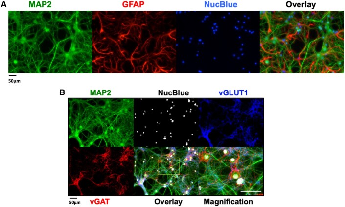
Mouse hippocampal neuron and glia co-culture at 14 days in vitro (DIV) express both glutamatergic and GABAergic neurons. A, Representative immunocytofluorescence images showing the neuronal/glial cultures at 14 DIV used to perform functional studies. Neuronal soma, axons and dendritic processes were stained with MAP2 and glia stained with GFAP. Nuclei were stained with NucBlue. Right-most panel is an overlay of all 3 stains. B, Representative immunocytofluorescence images showing the distribution of vesicular GABA transporters (vGAT) and vesicular glutamate transporters within MAP2 positive neurons. The overlay at higher resolution resolves the punctate pattern of the vGAT positive GABAergic neurons.

