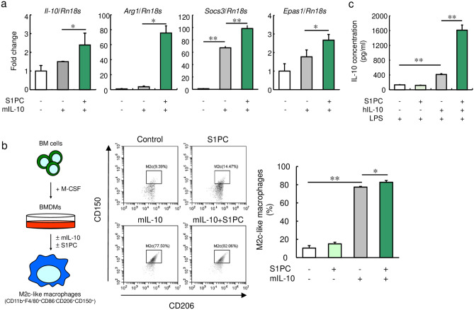
Figure 2.
Effect of S1PC on IL-10-induced macrophage polarization in M-CSF-induced BMDMs. (a) M-CSF-induced BMDMs were treated with S1PC (300 µM) in the presence of mIL-10 (20 ng/mL) for 48 h. The relative levels of M2 macrophage marker genes (Il-10, Arg1, Socs3, and Epas1) were analyzed using qRT-PCR. (b) M-CSF-induced BMDMs were treated with S1PC (300 µM) in the presence or absence of mIL-10 (20 ng/mL) for 48 h. The population of M2c-like macrophages (CD11b+, F4/80+, CD86-, CD206+, and CD150+ cells) were analyzed by flow cytometry. (c) M-CSF-induced BMDMs were treated with S1PC (300 µM) in the presence or absence of hIL-10 (20 ng/mL) for 48 h. Then, they were washed with PBS and treated with LPS (50 ng/mL) for 6 h. The amounts of IL-10 secreted into the culture media were determined using ELISA. Data are shown as mean + SD. Data are representative of three independent experiments. Statistical differences were determined using Bonferroni’s multiple comparison test (*p < 0.05 and **p < 0.01).

