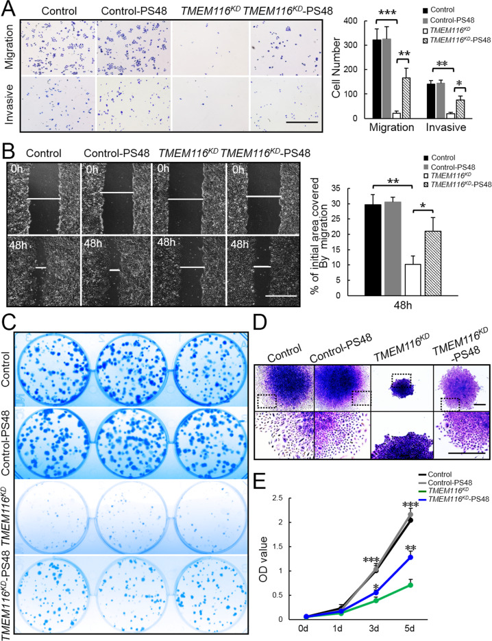
Fig. 6. Activation of PDK1 largely restores the proliferation, clone formation, migration and invasion of TMEM116KD cells.
A Control, control-PS48, TMEM116KD and TMEM116KD-PS48 cells were subjected to Transwell migration and invasion assays. More than three fields of cells in the lower chambers were counted. Scale bar: 1000 μm. B Control, control-PS48, TMEM116KD and TMEM116KD-PS48 cells were subjected to wound-healing assay. Scale bar: 1000 μm. Representative images from over 30 non-overlapping fields at each time point are shown. C Control, control-PS48, TMEM116KD and TMEM116KD-PS48 cells were subjected to colony formation assay. D Control, control-PS48, TMEM116KD and TMEM116KD-PS48 cells were subjected to colony morphology analyses. Scale bar: 500μm. E Control, control-PS48, TMEM116KD and TMEM116KD-PS48 cells were subjected to CCK8 assays at 0, 1, 3, 5 days. The bars represent the mean ± SD. *P < 0.05, **P < 0.01, ***P < 0.001. n = 6 mice. Representative images from three independent experiments are shown above.

