
Figure 7 .
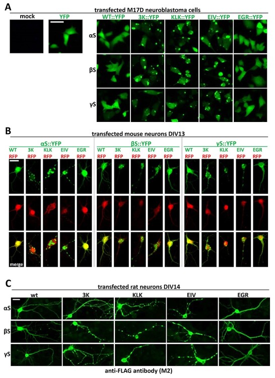
Fluorescence microscopy images of cells transfected with wt and variants of synuclein homologs. (A) M17D cells were transfected with YFP-tagged wt, 3K and KLK variants for all homologs. YFP was also transfected as a control. Images were taken 48 h post-transfection. Scale bar, 25 µm. (B) DIV 13 primary mouse neurons were co-transfected with YFP-tagged variants for all homologs and RFP as a control. Images were taken 48 h post-transfection. Scale bar, 20 µm. (C) DIV 14 rat neurons were transfected with YFP-tagged variants for all homologs. Images were taken 48 h post-transfection. All images are representative of N = 3 independent experiments done on different days. Scale bar, 20 µm.