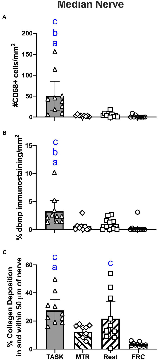
Figure 4.

Median nerve histomorphometry, at the level of the wrist. (A) Number of CD68+ macrophages. (B) Percent degraded myelin basic protein (dmbp) immunostaining in nerve. (C) Percent collagen deposition/fibrosis in and around the nerve in Masson’s Trichrome stained sections. Symbols: a, b, and c: p<0.05 each, compared to MTR, REST, and FRC groups, respectively. Mean±95% CI shown.
