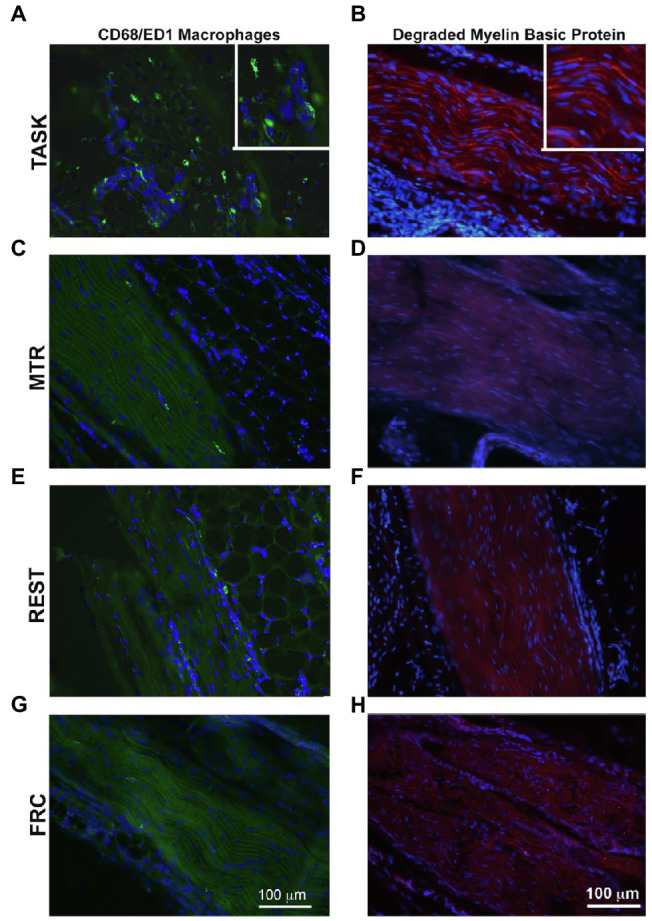
Figure 5.

CD68-immunopositive macrophages (green) and degraded myelin basic protein (red) in longitudinal sections of the median nerve. 4',6-diamidino-2-phenylindole (DAPI) was used as a counterstain (blue). (A,B) TASK rats. (C,D) MTR rats. (E,F) Rest rats. (G,H) FRC rats. Left panels: Representative images of CD68+ cells in median nerves at wrist level. Right panels: Representative images of degraded myelin basic protein in median nerves at wrist level. Scale bars in panels A and B=100μm and applies to the other panels.
