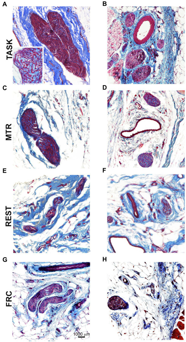
Figure 7.

Masson’s Trichrome staining for collagen in and around median nerves at wrist level. Left panels (A,C,E,G): Representative images of longitudinal sections of the median nerve. Right panels (B,D,F,H): Representative images of cross-sections of the median nerve. Scale bar in panel G=1,000μm and applies to the other panels.
