FIGURE 1.
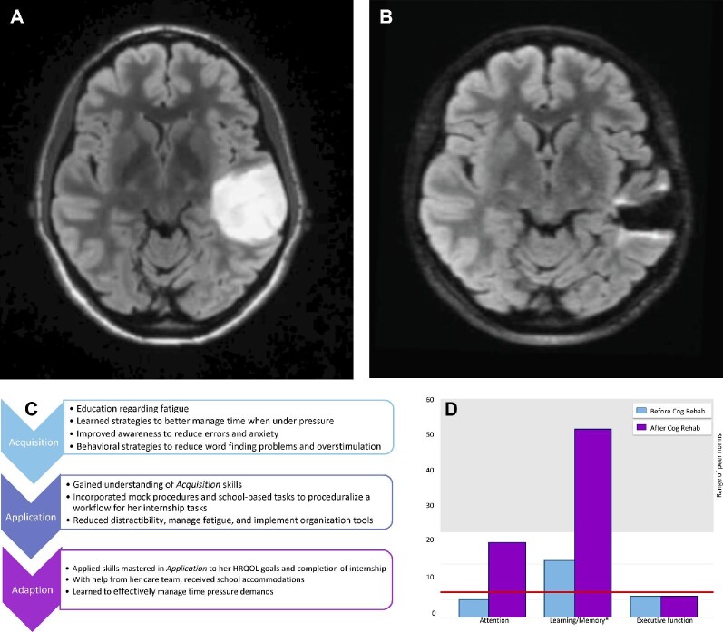
Case example of a 29-yr-old right-handed Caucasian woman who presented with a generalized seizure and 10 yr of word finding difficulty and forgetfulness, and was found to have a nonenhancing left temporal mass. A, Axial MRI demonstrating T2/FLAIR hyperintense mass in the left temporal lobe prior to initial surgery. She underwent gross total resection with intraoperative awake mapping with pathology confirming diffuse astrocytoma, IDH mutant, with anomia of low-frequency words and expressive dysphasia that improved with occupational and speech therapy. She underwent a second surgery 12 mo later and postoperatively developed distractibility, forgetfulness, and reduced multitasking. She was referred for neuropsychological evaluation, with symptoms negatively impacting her schoolwork. B, Axial MRI demonstrating minimal T2/FLAIR hyperintense lesion in the left temporal lobe at time of referral to cognitive rehabilitation. C, She underwent 8 sessions of cognitive rehabilitation via telehealth over 4 mo, using the Triple A model. D, Baseline testing (blue) revealing mild-to-moderate vulnerabilities in distractibility, thinking speed, word finding, verbal memory, and executive functioning. Postintervention testing (purple) revealed improved auditory attention, working memory, and verbal memory. The asterisk denotes clinical significance at one standard deviation. The red line is the cutoff for clinical impairment and the gray-shaded area depicts the range of adjusted peer normative values. She went on to successfully pass her internship and licensing exam, and at the end of treatment, was working part-time in her preferred field (presented with patient permission). FLAIR, fluid-attenuated inversion recovery.

