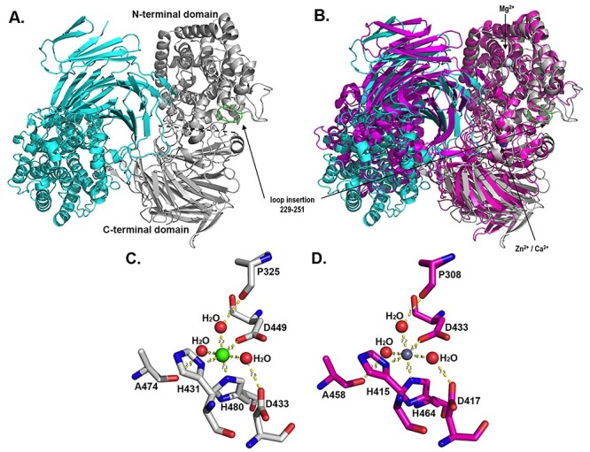
Fig. 5.

Crystal structure representation of the AlyA3 biological dimer. (A) Cartoon representation of the 2-fold dimeric quaternary arrangement of AlyA3. Monomer A is colored in cyan and monomer B in light gray. In the gray monomer, the N-terminal (α/α)6 toroid and a C-terminal β-sheet domain are separated by a dotted line. The loop insertion found in AlyA3 but absent in Alg17c (see Figure 1) is colored in green and indicated by an arrow. (B) Superimposition of the dimer of AlyA3 (cyan and gray) with the dimer of Alg17c (colored in magenta, pdb code 4OJZ). The positions of ions in AlyA3 are indicated by an arrow. (C) Stick representation of the conserved residues together with the water molecules forming the octahedral coordination sphere of the Ca2+ ion in AlyA3. (D) Stick representation of the conserved residues together with the water molecules forming the octahedral coordination sphere of the Zn2+ ion in Alg17c. The figure was produced using PyMOL (The PyMOL Molecular Graphic Systems 2002). This figure is available in black and white in print and in color at Glycobiology online.
