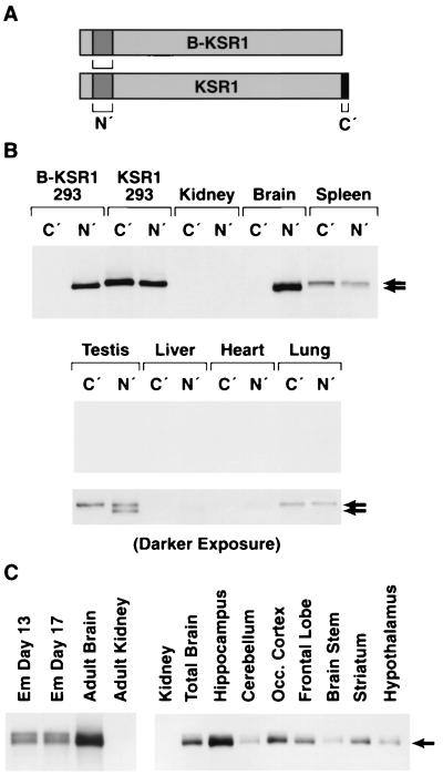
FIG. 2.
Expression of B-KSR1 and KSR1 proteins in various mouse tissues. (A) Immunoprecipitation assays were performed using two antibodies, αN′ (N′) and αC′ (C′). The presence of these epitopes on B-KSR1 and KSR1 is depicted. (B) Lysates prepared from 293 cells expressing either B-KSR1 or KSR1 and lysates from various adult mouse tissues were equalized for protein content and immunoprecipitated with either αN′ or αC′. The immunoprecipitates were then examined by immunoblot analysis using αN′. Short and long exposures are shown. (C) Kidney and brain tissue from adult mice, brain tissue from mice at embryonic (Em) days 13 and 17, and tissues from specialized brain regions were isolated, and lysates were prepared. The lysates were equalized for protein content and immunoprecipitated with αN′. The immunoprecipitates were then examined by immunoblot analysis using an antibody recognizing KSR.

