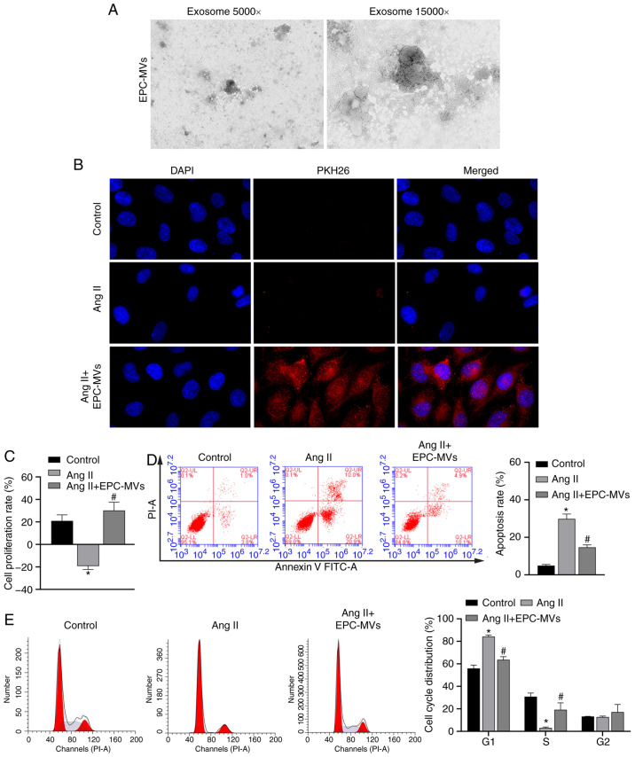
Figure 3.
Recovery of PRKs by co-culture with EPC-MVs following injury induced by Ang II. (A) Successful isolation of EPC-MVs from EPCs as determined via transmission electron microscopy. Magnification, ×5,000 and ×15,000. (B) Fusion of PKH26- labeled EPC-MVs with PRKs, Magnification, ×400. (C) CCK-8 analysis of the effects of different proportions of co-cultured EPC-MV on the proliferation of PRKs. Flow cytometry analysis of the effects of co-cultured EPC-MVs on the (D) apoptosis and (E) cell cycle of Ang II-treated PRKs. *P<0.05 vs. Control; #P<0.05 vs. Ang II. Ang II, angiotensin II; EPCs, endothelial progenitor cells; PRKs, primary rat kidney cells; MVs, microvesicles.

