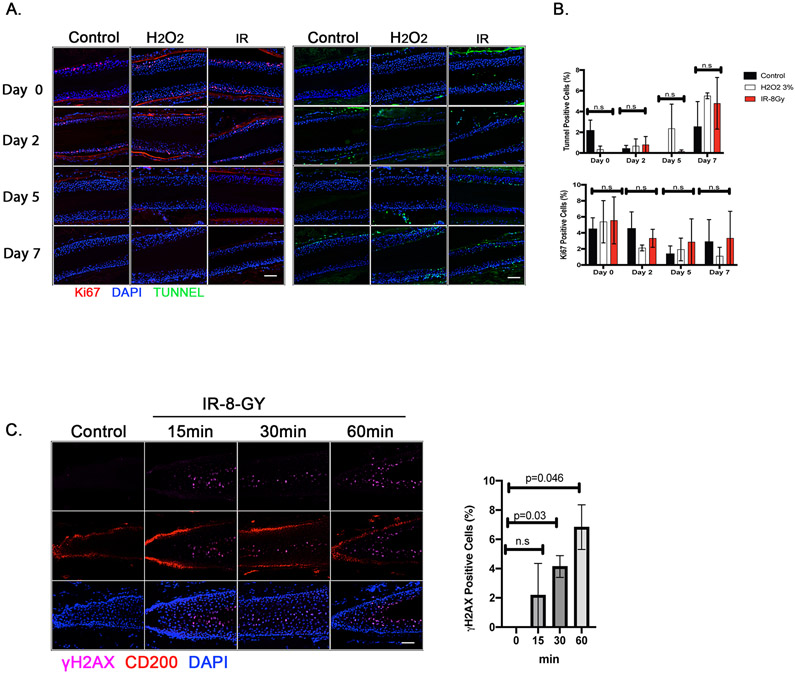
Figure 2. Effect of genotoxic stress (hydrogen peroxide and ionizing radiation) on viability and DNA damage in isolated human hair follicles.
Human hair follicles (HFs) were isolated and irradiated (8 Gy) or treated with 3% hydrogen peroxide. A. Viability was tested at days 0, 2, 5 and 7 post treatment using immunofluorescent staining for Ki-67 (a proliferation marker; red) and TUNNEL assay (apoptosis; green) in the ORS (Scale bar; 50μm). B. Ki-67-and TUNNEL-positive cells were quantified and normalized to DAPI-positive ORS cell counts. Data are mean ± standard error of mean (SEM). n = 2 human donors; 48-50 hair follicles from each donor were analyzed; n.s., not significant; p values were determined by two-way ANOVA. C. Immunofluorescent staining of hair follicles for the DNA damage marker (γ-H2AX; purple), for hair follicle at the bulge (CD200; red), and for nuclei (DAPI). Scale bar, 50μm. The percentage of γ-H2AX-positive cells were calculated out of the total DAPI-positive cells at the bulge area (CD200-positive area). Data are mean ± SEM. 48-50 hair follicles were analyzed from each of 2 independent donors. p values were calculated by the two-tailed Student’s t-test; p < 0.05 is considered statistically significant.

