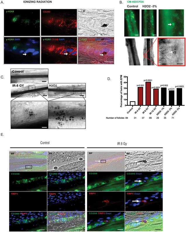
Figure 3. Induction of ectopic pigmentation by IR and H2O2 in the niche of human HFs.
A. Human HFs were isolated and irradiated (8 Gy). The pigmented cells are observable under brightfield microscopy (black spots). Human HFs were immunolabeled with CD200 (bulge; red) and γH2AX (a DNA damage maker; green). Scale bar, 100μm. The pigmentated cells are indicated by white Arrow. B. Isolated human HFs were treated with 3% H2O2 for 2 hours. ROS-positive cells were immunolabeled with CM-H2DCFDA (green) at the bulge area of the hair follicles. The black-pigmented cells appear around the ROS-positive cells (white arrow). White line-scale bar, 20μm; black line-scale bar, 100μm C. Representative follicle showing ectopic pigmentation after IR and H2O2 treatment. EPM, ectopically pigmented melanocytes. Black line-scale bar, 20μm; white line-scale bar, 100μm. D. Quantification of pigmentation efficiency (percentage of HFs with EPM divided by the total number of HFs) 3 days post treatment. n = 8 human donors, 29 to 99 hair follicles. p values were calculated by the Fisher’s exact test; p < 0.05 is considered statistically significant. E. Immunofluorescent staining of TRP-1 (red) for human HFs after IR (8 Gy). Arrow indicates ectopic pigmentation co-localized with TRP-1 staining.

