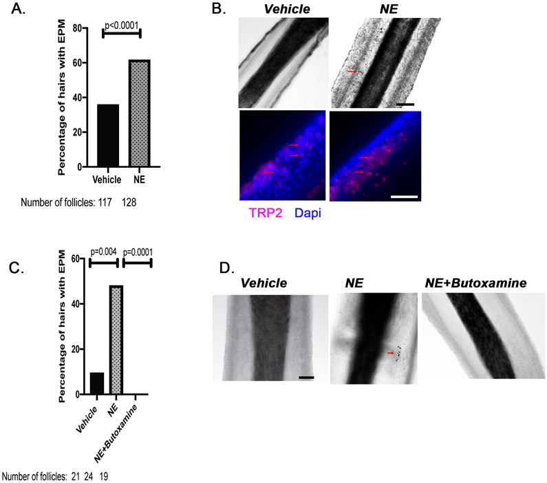
Figure 4. Noradrenaline induces ectopic pigmentation in the ORS, which can be prevented by blocking the β-2 adrenergic receptor.
A. Human HFs were isolated and treated with 0.1mM noradrenaline, or vehicle (deionized distilled water) for 24 hours. n = 2 different human donors, 117 to 128 hair follicles. p value was calculated by the Fisher’s exact test; p < 0.05 is considered statistically significant. B. Immunofluorescence staining of hair follicles for the melanocytic marker, TRP-2 (Red). Brightfield images of the isolated hair follicles with ectopic pigmentation observed as black cells as indicated by red arrows. Scale bar, 20μm. C. Isolated human HFs were treated with vehicle (deionized distilled water), noradrenalin (0.1 mM), or noradrenalin (0.1 mM) + butoxamine (0.1 mM) for 24 hours. EPM, ectopically pigmented melanocytes. n = 1 human donor, 19 to 24 hair follicles. p values were calculated by the Fisher’s exact test; p < 0.05 is considered statistically significant. D. Representative brightfield images of the isolated HFs. Scale bar, 20μm.

