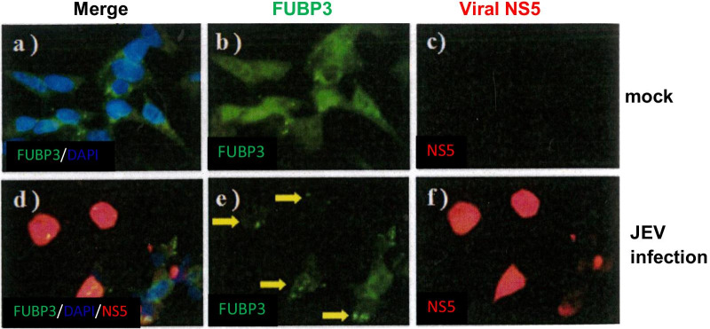
Fig. 4.
Detection of colocalization of FUBP3 with JEV-NS5 protein in JEV-infected BHK-21 cells. Mock- or JEV-infected BHK-21 cells were harvested at 48 h post-infection and prepared for immunofluorescence analysis stained with antibodies that detect FUBP3 (green) and JEV-NS5 protein (red). Subcellular localization of FUBP3 (panel b, e) and viral NS5 protein (panel f) in mock- and JEV-infected cells. The nucleus was stained with DAPI as shown in the merged image (panel a, c). Arrows showed the colocalization of FUBP3 with NS protein (panel e) were detected in the cytoplasm of JEV-infected cells. The Pearson correlation coefficient (PCC) of images of FITC-FUBP3 and Texas Red-NS5 is 0.9998. Images are representative of three independent experiments that used three independent infections

