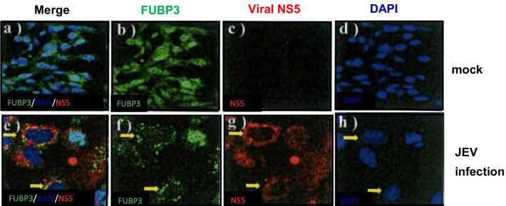
Fig. 5.
Overexpression of FUBP3 colocalized with viral NS5 protein in JEV-infected BHK-21 cells. Flag-tagged FUBP3 protein that was expressed in BHK-21 cells 24 h prior to JEV infection. The mock- and JEV-infected BHK-21 cells were harvest at 48 h post-infection and co-immunostained with anti-NS5 (red) and anti-FUBP3 (green) antibodies. Subcellular localization of FUBP3 (panel b, f) and viral NS5 protein (panel g) in mock- and JEV-infected cells. The nucleus was stained with DAPI as shown in the merged images (panel a/d, e/h). Arrows showed the colocalization of FUBP3 with NS protein (panel e) in the cytoplasm of JEV-infected cells. The Pearson correlation coefficient (PCC) of images of FITC-FUBP3 and Texas Red-NS5 is 0.9946. Images are representative of three independent experiments that used three independent infections

