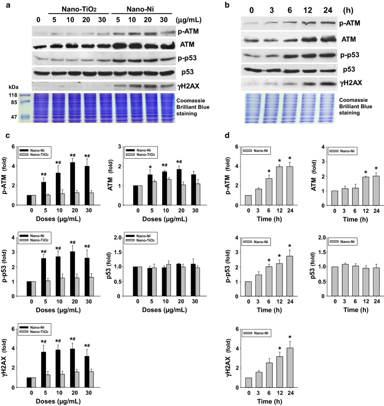
Fig. 2.
Nano-Ni exposure caused increased expression of DNA damage response-associated proteins in BEAS-2B cells (dose- and time-response studies). For the dose–response study, cells were treated with 5, 10, 20, and 30 µg/mL of Nano-Ni or Nano-TiO2 for 24 h. For the time-response study, cells were treated with 20 µg/mL of Nano-Ni for 3, 6, 12, and 24 h. Cells without treatment were used as the control. Nuclear protein was subjected to Western blot. Equal nuclear protein loading was verified by Coomassie Brilliant Blue staining. A, B are results of a single Western blot experiment, while C, D are quantified band densitometry readings averaged from at least 3 independent experiments ± SEM of Western blot results. * p < 0.05 vs. control; # p < 0.05 vs. same dose of Nano-TiO2-treated group. p-ATM, phosphorylated ATM at Ser1981; p-p53, phosphorylated p53 at Ser15

