Fig. 6.
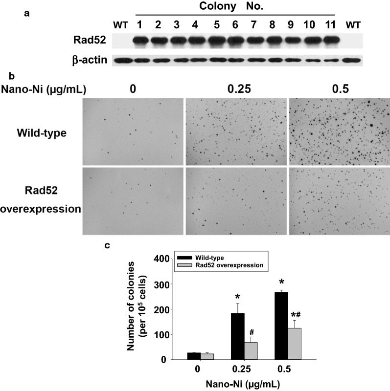
Long-term Nano-Ni exposure induced cell transformation, which was attenuated by overexpression of Rad52. A BEAS-2B cells were transduced with lentiviral particles containing human Rad52 ORF as described in the Methods. 11 puromycin-resistant colonies were picked and expanded, and the total protein from each colony was isolated for Western blot. The exposure time was 2 s. β-actin served as loading control. WT, wild-type. B, C The effects of Nano-Ni on anchorage-independent growth of cells by soft agar colony formation assay. Cells were exposed to 0, 0.25 and 0.5 µg/mL of Nano-Ni for 21 cycles as described in the Methods. The colonies were stained with INT/BCIP solution (B) and quantified using ImageJ software (C). Data are shown as mean ± SEM (n = 3 ~ 6). *, p < 0.05 vs. control; #, p < 0.05 vs. wild-type group with same dose of Nano-Ni treatment

