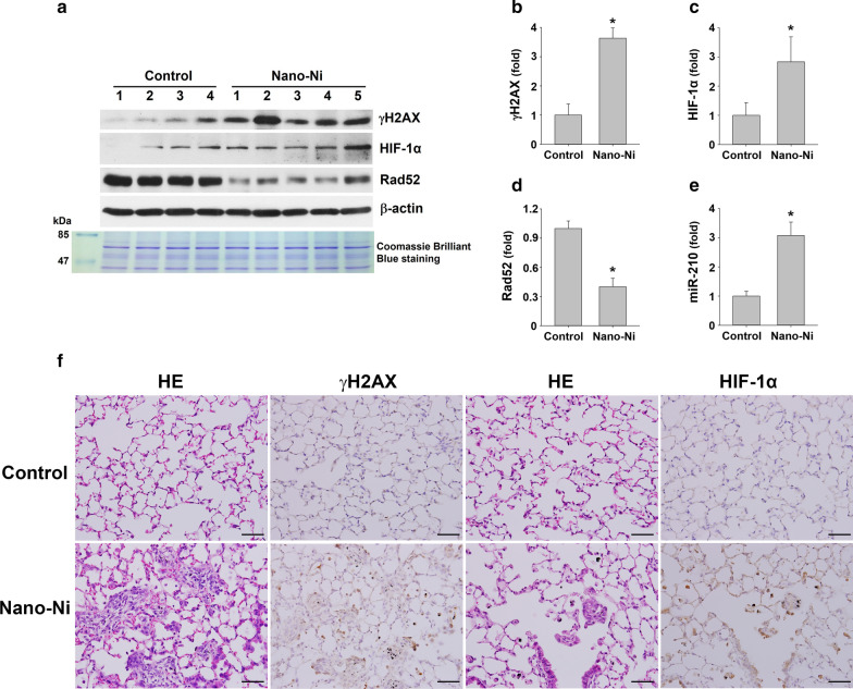
Fig. 7.
HIF-1α/miR-210/Rad52 signaling and expression of DNA damage response protein in mouse lungs after Nano-Ni exposure. Mice were instilled intratracheally with 50 µg per mouse of Nano-Ni. Control mice were instilled with physiological saline. Lung tissues were collected at day 7 after Nano-Ni exposure. A is the results of Western blot experiment, while B–D are results quantified by ImageJ software and normalized by internal control β-actin. E miR-210 expression was determined by real-time PCR. Values of miR-210 expression was normalized to the endogenous control U6 snRNA. Data are shown as mean ± SEM (n = 4–5). *, p < 0.05 vs. control. F Expression of HIF-1α and γH2AX in mouse lungs by immunohistochemical staining. Increased number of HIF-1α and γH2AX positive cells (brown staining) were observed in the mouse lungs after Nano-Ni exposure. Scale bars represent 50 µm for all panels

