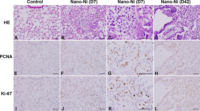
Fig. 8.
Increased number of PCNA- and Ki-67-positive cells in mouse lungs after Nano-Ni exposure by immunohistochemical staining. Mice were instilled intratracheally with 50 µg per mouse of Nano-Ni. Control mice were instilled with physiological saline. Lung tissues were collected at day 7 (D7) and day 42 (D42) after Nano-Ni exposure. A, E, and I show the normal structure of lung parenchyma in a control mouse. Increased number of PCNA (F–H) and Ki-67 (J-L) positive cells (brown staining) were observed in the mouse lungs after Nano-Ni exposure. Scale bars represent 50 µm for all panels

