
Figure 2.
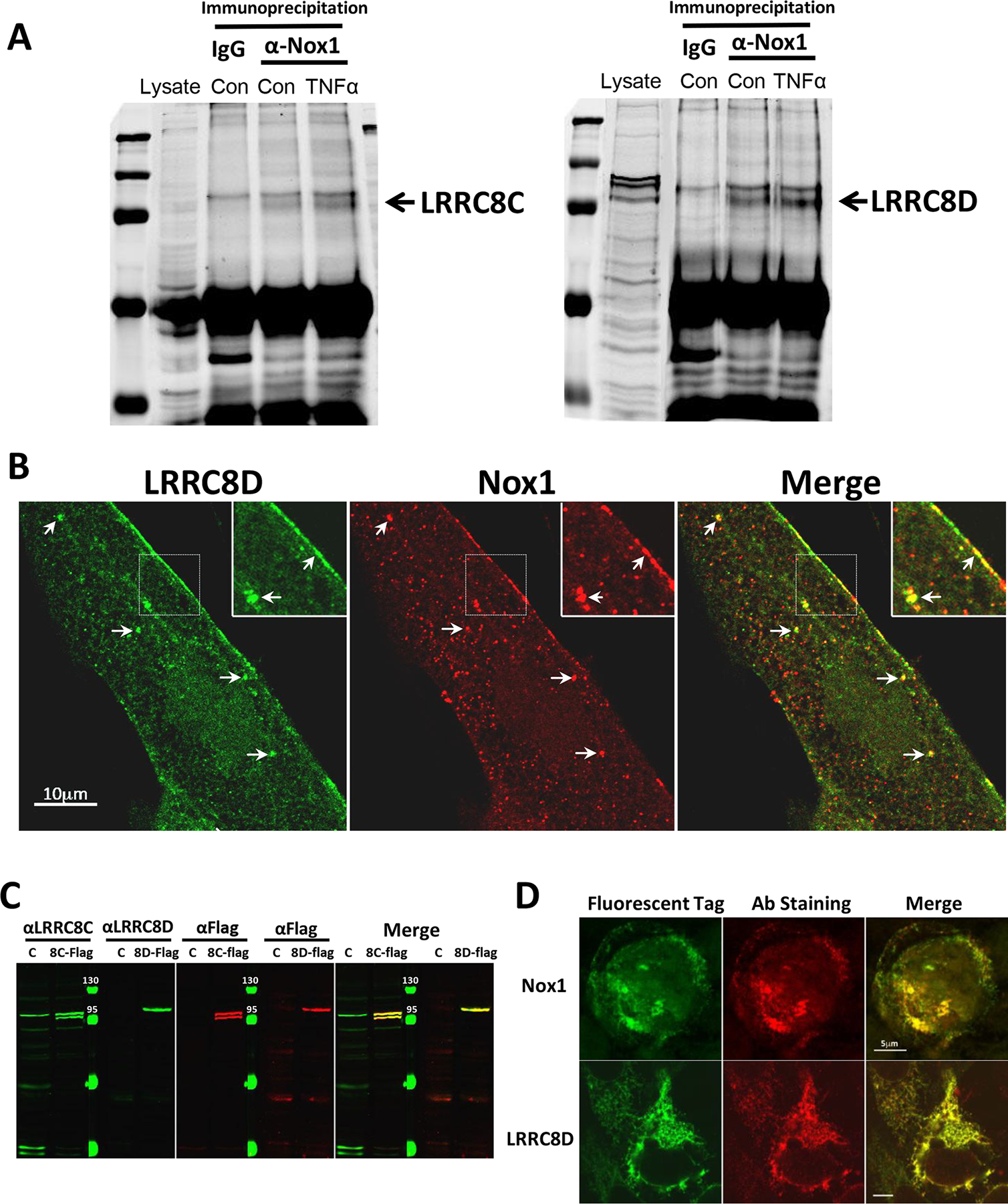
A, Control IgG or anti-Nox1 Ab were used to immunoprecipitate proteins from cultured human VSMC cell lysates under control conditions or following a 10 min exposure to TNFα. Western blotting for LRRC8C (left) or LRRC8D (right) proteins was performed on whole cell lysates (Lysate) or the immunoprecipitated proteins. Anti-Nox1 pulled down both LRRC8 proteins but this association was not enhanced by exposure to TNFα (n = 3). B, Immunostaining of cultured human VSMCs with anti-LRRC8D and anti-Nox1 reveals co-localization both at the plasma membrane and in intracellular vesicles (arrows; insets reflect 2X magnification). C, Western blots of protein obtained from Control HEK293 cells (C), or HEK cells expressing flag-tagged LRRC8C (8C-Flag, left) or LRRC8D (8D-Flag, right). Blots were probed using either αLRRC8C or αLRRC8D and αFlag and imaged using the Odyssey Imaging System. The protein-specific (green) and αFlag antibodies (red) identified the same bands in expressing cells (yellow signal in Merged image). αLRRC8C identified a band in the Control lysate of the same size as expressed LRRC8C but LRRC8D was not identified in the HEK293 cell lysate. D, HEK293 cells heterologously expressing GFP-tagged Nox1 (top) or LRRC8D (bottom) were immunostained with anti-LRRC8D or anti-Nox1 (red, middle panels) to validate antibody specificity. GFP and antibody labeling were strongly colocalized for both proteins (yellow signal, Merge panels).