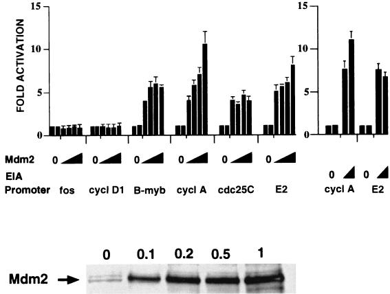
FIG. 1.
Activation of cell cycle-responsive promoters by Mdm2 expression in tsBN462 cells. The cells were transfected in six-well dishes with luciferase reporters containing promoters from the fos, cyclin D1 (cycl D1), B-myb, cyclin A (cycl A), cdc25C, and adenovirus E2 genes (1 μg) and increasing amounts of expression vectors for Mdm2 (pCMV-Mdm2; 0.1, 0.2, 0.5, and 1 μg) or E1A (0.5 and 1 μg). After transfection the cells were transferred to 39°C, and 24 h later the luciferase activities were measured. Luciferase activities were corrected for variations in transfection efficiency by using the internal control (pSG5-LacZ; 1 μg/ml). They are presented relative to the control (set to 1) that was included in duplicate in each experiment. The fold activations are the averages from three independent experiments, and the error bars indicate the standard deviations. The Western blot in the lower panel was probed with rabbit anti-Mdm2 antibody no. 365 raised against a mouse Mdm2-derived peptide (33). The arrow indicates the Mdm2-specific band migrating at a molecular weight of around 90,000. The lane markers 0, 0.1, 0.2, 0.5, and 1 correspond to the amounts (in micrograms) of transfected pCMV-Mdm2.

