FIG. 7.
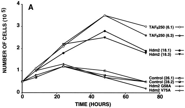
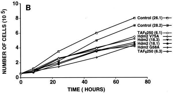
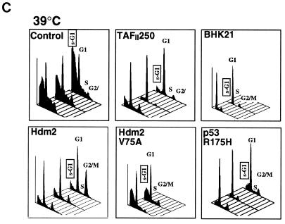
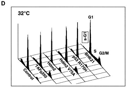
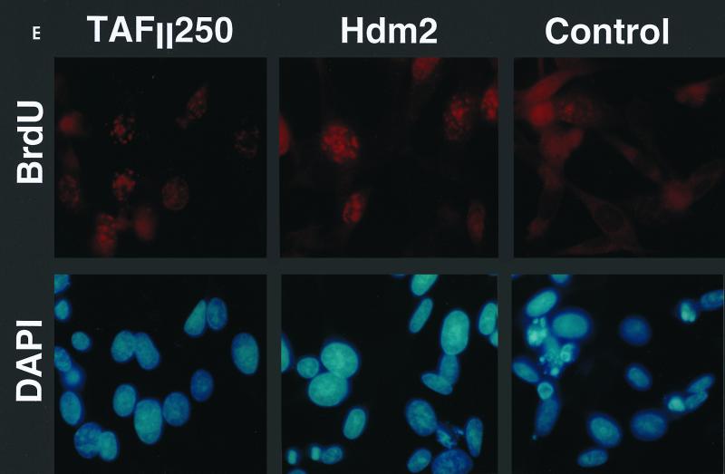
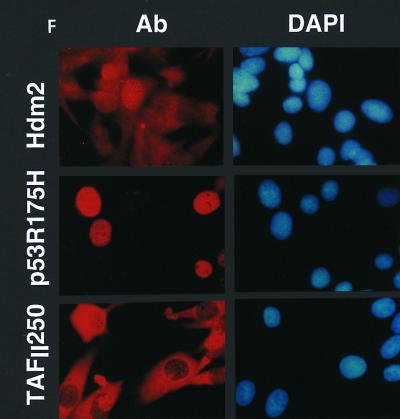
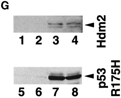
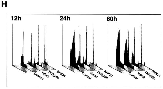
Effect of TAFII250, Hdm2, and p53 R175H expression on cell growth, cell cycle distribution, DNA synthesis, and long-term survival. Stable G418-resistant clones were established that express TAFII250, Hdm2, Hdm2 G58A, Hdm2 V75A, or p53 R175H or contain the empty pCMV expression vector. (A and B) The cells were plated in six-well dishes (5 × 10⁴ per well) and incubated at 32°C for 2 h before being incubated at either 39°C (A) or 32°C (B). At the indicated times, the cells were collected by trypsinization and viable cells (that exclude trypan blue) were counted in a Bürker cell. The clone numbers are indicated in parentheses. (C and D) Exponentially growing cells were incubated at 39°C (C) or 32°C (D) for 24 h and analyzed by flow cytometry. The scans for independent clones are shown in panel C (Control, clones 26.1, 28.2, and 28.3; TAFII250, clones 6.1, 6.2, and 6.3; BHK21, two plates analyzed separately; Hdm2, clones 18.1, 18.2, and 18.3; Hdm2 V75A, clones 24.1 and 23.1; p53 R175H, clones 12.1, 12.2, and 11.1). One representative clone for each vector is shown in panel D. s-G1, sub-G0/G1 (boxed and rotated); G1, G0/G1. (E) The clones were incubated for 12 h at 39°C, labeled with BrdU for 1 h, fixed, and incubated with anti-BrdU (Becton-Dickinson) and Texas red anti-mouse (Jackson) antibodies, and DAPI. Fluorescent cells in 10 different fields containing 10 to 50 cells were counted. The proportions of BrdU-positive cells were as follows: TAFII250 (clones 6.1 and 6.3), 33% ± 5%; Hdm2 (clones 17.2 and 18.2), 35% ± 4%; Control (clones 27.2 and 28.3), 4% ± 2%. Representative photographs are shown. (F) Cells were fixed, stained with specific antibodies (Hdm2, IF2; p53R175H, DO1; TAFII250, anti-HA, 12CA5) followed by Cy3-labeled secondary antibodies and DAPI (nuclei). The cells were photographed under a fluorescence microscope. Control clones incubated with the specific antibodies and labeled secondary antibodies gave low levels of background fluorescence (not shown), as expected for antibodies that are specific for human or viral proteins. One representative clone for each is shown. (G) Western blots of clones established with the selectable marker alone (lanes 1, 2, 5, and 6) or with expression vectors for Hdm2 (lanes 3 and 4) or p53R175H (lanes 7 and 8) and probed with antibodies against Hdm2 (Ab-1, Calbiochem OP46 [lanes 1 to 4]) or p53 (DO1 [lanes 5 to 8]) followed by enhanced chemiluminescence (ECL) (Amersham). (H) Exponentially growing cells were incubated at 39°C for 12, 24, and 60 h and analyzed by flow cytometry. One representative clone for each vector is shown.

