FIG. 8.
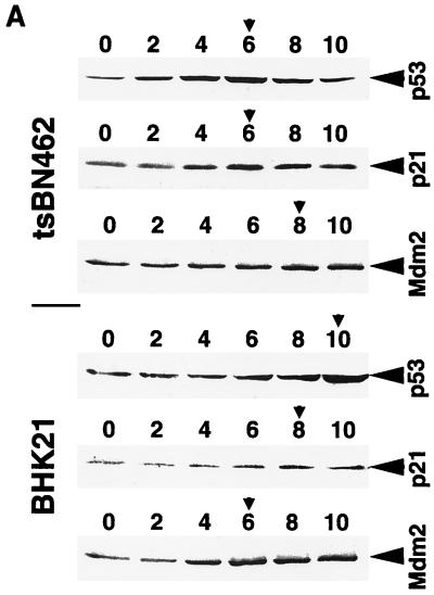
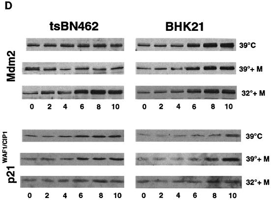
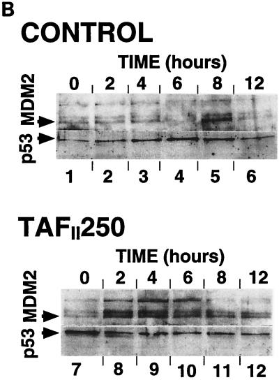
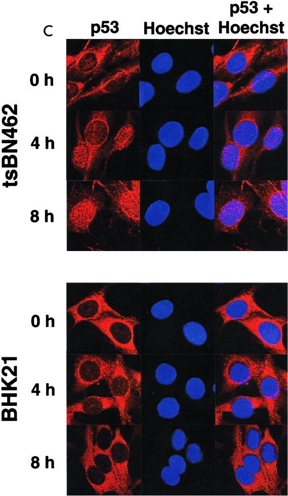
p53, p21WAF1/CIP1, and Mdm2 protein expression in tsBN462 and BHK21. (A) Exponentially growing tsBN462 and BHK21 cells were transferred to 39°C. At the indicated times (0, 2, 4, 6, 8, and 10 h), cell extracts were prepared by lysis in loading buffer and analyzed by SDS-PAGE (8% polyacrylamide), Western blotting, and ECL (Amersham), as described previously (33). The antibodies used were as follows: p53, PAb 240; p21WAF1/CIP1, C-19-G (sc-397-G [Santa Cruz]); Mdm2, 2A10 (similar results were obtained with SMP14 [data not shown]). Arrows pointing to the left indicate specific bands with the expected mobility. Arrowheads pointing down indicate the time point with the maximum level of expression. Ponceau S staining of the membranes and blotting for RNA polymerase II large subunit (a gift from M. Vigneron) were used to verify equal loading (not shown). (B) tsBN462 cells were transfected in 9-cm dishes with vectors that express HOOK (1 μg pHOOKTM-1) and either TAFII250 (10 μg) or nothing (Control) (10 μg). Transfected cells were isolated with the Capture-Tec kit (Invitrogen), replated, cultivated for 18 h, and then induced by heat shock at 39°C. Samples were collected at different times (0, 2, 4, 6, 8, and 12 h) and analyzed by SDS-PAGE (8% polyacrylamide), Western blotting, and ECL to detect Mdm2 (SMP14) and p53 (DO1). Ponceau S staining of the membranes was used to verify equal loading (not shown). (C) tsBN462 and BHK21 cells were transferred to 39°C, and at different times (0, 4, and 8 h) the cells were fixed, stained, and analyzed by confocal microscopy, as described previously (63). p53 was revealed with a rabbit antibody (no. 588) followed by Cy3-labeled goat anti-rabbit antibody (Jackson). Similar localizations were observed with PAb 240. Hoechst stains nuclei. Typical fields are shown. (D) Exponentially growing tsBN462 and BHK21 cells were incubated at 39°C or 32°C in the absence or presence (+M) of mitomycin C (5 μg/ml). At the indicated times (0, 2, 4, 6, 8, and 10 h), cell extracts were analyzed by SDS-PAGE (8% polyacrylamide) and Western blotting (antibodies: p21WAF1/CIP1, C-19-G [sc-397-G]; Mdm2, 2A10) and ECL. Protein quantification (Bio-Rad protein assay), Ponceau S staining of the membranes, and blotting for RNA polymerase II large subunit (a gift from M. Vigneron) were used to verify equal loading (not shown).


